Unit of Forensic Imaging (UIF)
PRESENTATION | SERVICES | RESEARCH & TRAINING | FAQ
Presentation
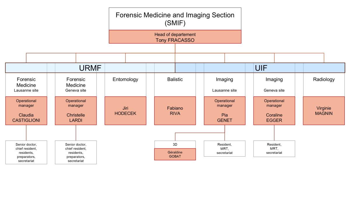
The Unit of Forensic Imaging is part of the Forensic Medicine and Imaging Section (SMIF)
Main tasks
- The UIF performs at the request of the judicial authorities forensic imaging investigations, a technique that includes methods for acquiring and restoring images of the human body taking advantage of various techniques of imaging (e.g. CT-scan and MRI). Imaging investigations precede and complement the autopsy. Imaging techniques have the advantage of being non-destructive and the acquired data can be stored and reviewed later on.
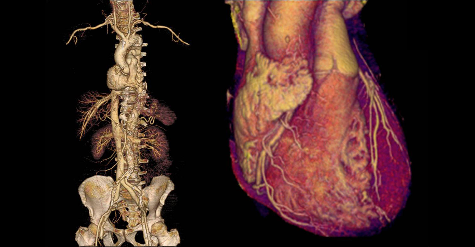
- Postmortem angio-CT is a minimally invasive imaging method that allows to investigate the details of the vascular system that can't be achieved with a conventional autopsy. This examination requires the administration of a contrast agent with an infusion pump. CT angiography enables the detection of a source of bleeding, a malformation of the network of blood vessels, arteriosclerosis lesions, occlusion of a vessel and the visualization of the vascular anatomy.
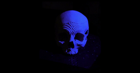
- 3D documentation which allows reconstruction by virtual 3D modelling and 3D morphometric comparisons between the lesions and the suspected objects of causing the lesions.
- For enquiries, please contact our secretary : Melissa Jotterand
Field of activity
The main fields of activity deal with postmortem investigation, clinical expertise and expertise on record. The UIF also provides teaching and training to under-graduate and post-graduate students and also in continuing education. A wide variety of professions may be interestedv: medical doctors, pathologists, scientists, paramedics, judges, lawyers, police,...). The UIF develops innovative research programmes in the field of forensic imaging.
Staff
- The activities of the UIF at the Geneva and Lausanne sites are carried out under the responsibility of Prof. Dr. Tony Fracasso. The unit is composed of forensic doctors, radiologists, radiographers, secretaries and specialists in 3D surface scanning.
| UIF |
|
|---|---|
| Staff | |
| Head of the unit | Tony Fracasso |
| Forensic pathologist |
Coraline Egger (GE) Pia Genet (LS) |
| Radiologist | |
| Medical trainee |
Javan Helbling (GE) |
| TRM | Alejandro Dominguez (LS) |
| Christine Bruguier (LS) | |
| Ruben Soto (GE) | |
| Sami Schranz (GE) | |
| Vadim Udhe (GE) | |
| Valerie Frossard (LS & GE) | |
| Lucia Fernandes Mendes (LS) | |
| Dellaram Anissi (LS) | |
| 3D surface scanning specialist | Géraldine Gobat |
| Ballistics expert | Fabiano Riva |
| Secretary | Melissa Jotterand (LS) Annick Crockett-Griessen (GE) Agnès Chapuis (LS) |
| Consulting doctors | external radiologists |
The UIF also welcomes interns and visiting researchers for varying periods of time, including students (medical, radiographer), foreign doctors, radiographers who wish to specialise in forensic imaging.
Equipments
Lausanne site:
- Radiological imaging equipments (Mobile device Agfa DX-D100, CT-Scan (GE HealthCare CT Discovery HD 750, 64-rows)
- Virtangio machine (Postmortem injector)
- 3D surface scanner GOM ATOS Compact Scan M5 (Camera MV150 et MV300)
- One handheld 3D surface scanner iReal M3.
- MRI Philips Ingenia CX 1,5T.
Geneva site:
- Radiological imaging equipments (Mobile device Agfa DX-D100, CT-Scan (GE HealthCare LightSpeed VCT 64 barrettes)
- Virtangio machine (Postmortem injector)
- Two handheld 3D surface scanner CREAFORM Go!SCAN 50 et Go!SCAN 20.
- 3D Printer (3D-Systems ZPrinter 150)